|
Packaging Details - Unit Type:piece
- Package Weight:0.3kg (0.66lb.)
- Package Size:30cm x 20cm x 20cm (11.81in x 7.87in x 7.87in)
Packaging Details - Unit Type:piece
- Package Weight:0.3kg (0.66lb.)
- Package Size:30cm x 20cm x 20cm (11.81in x 7.87in x 7.87in)
tyle="max-width:650px;overflow:hidden;font-size:0;clear:both"> /piece /piece /piece /piece /piece /piece /piece /piece Product model: ESZXT023 Product size: 26*11*11cm Package size: 30*20*20cm Packing weight: 0.3KG ENOVO new testes model testicular structure reproductive and urological male reproductive health guidance model The testis is located in the scrotum, about one or so, and the left side is slightly lower than the right 1cm. The testes are slightly oval, with smooth surfaces, two sides of the inside and outside sides, the front, rear, and upper and lower ends. The anterior margin is free, and the posterior edge is surrounded by blood vessels, nerves and lymphatics, and is in contact with the testis of the epididymis and the vas deferens. The upper and posterior margins are epididymis and attached to the lower end. The lateral surface is more convex, and the inner surface is flat. The structure of the testis The fibrous membrane, called the white membrane, thickens the white membrane along the posterior edge of the testis, and enters the testicle to form the mediastinum of the testis. From the mediastinum, many connective tissue septum is issued and the testicular parenchyma is divided into many testicular lobules. The testicular lobules contain convoluted seminiferous tubules, and the epithelium of the seminiferous tubules can produce spermatozoa. There are interstitial cells secreting male hormones in connective tissue between tubules. The seminiferous tubules are combined into seminiferous tubules, and into the testis mediastinum interwoven into the testis web. From testis net, 12~15 testicular output tubules and the upper part of the posterior edge of testis enter the epididymis. The model was designed to amplify 3.5 times normal male testicles to show the inside and sagittal section of the testis, and the internal structure of the testis was displayed in detail, such as the output of the tubules, the white film, the vaginalis, the vaginalis, the spermatogonia, the vas deferens, the spermatic duct and the testicular net. It is a rare product in the teaching of reproductive science and male families. It is the main appliance for the communication between doctors and patients at present, and it is also suitable for the teaching demonstration in the hospital. 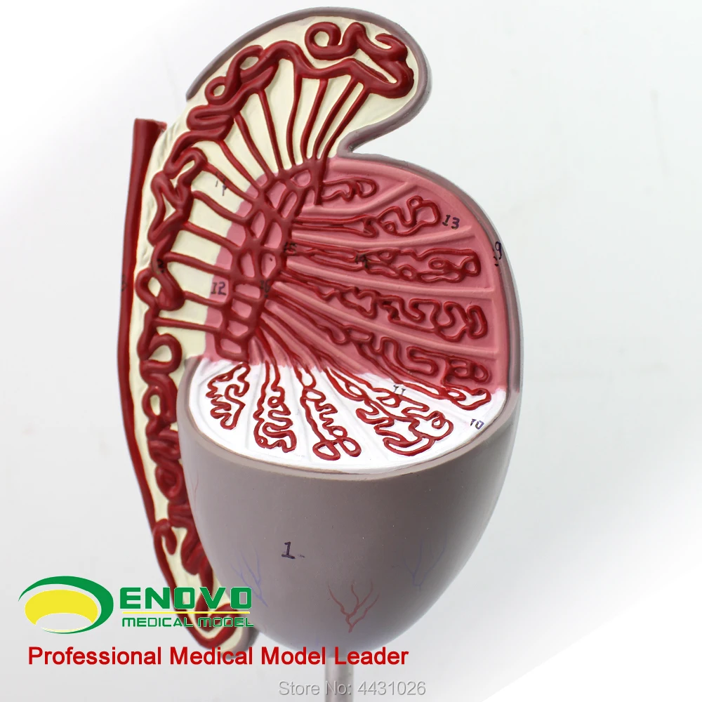 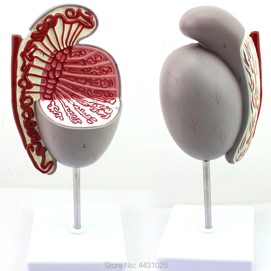 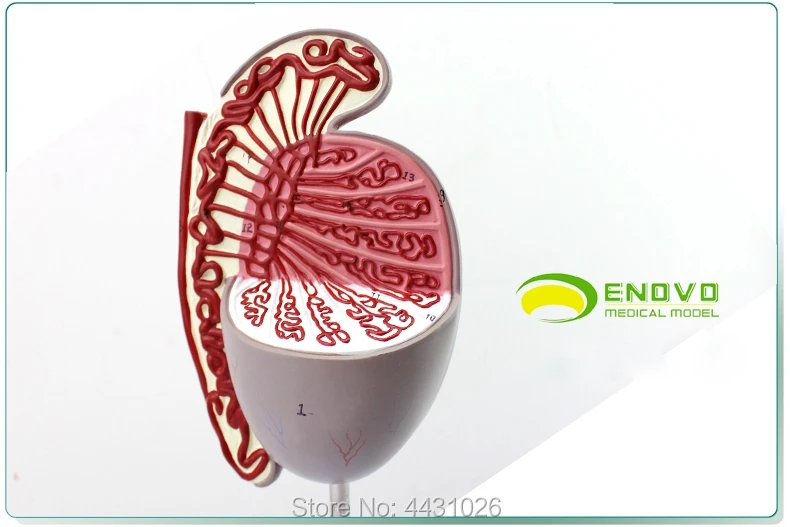   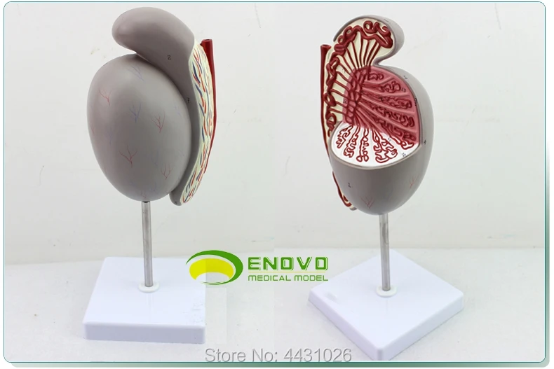 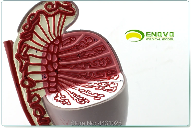 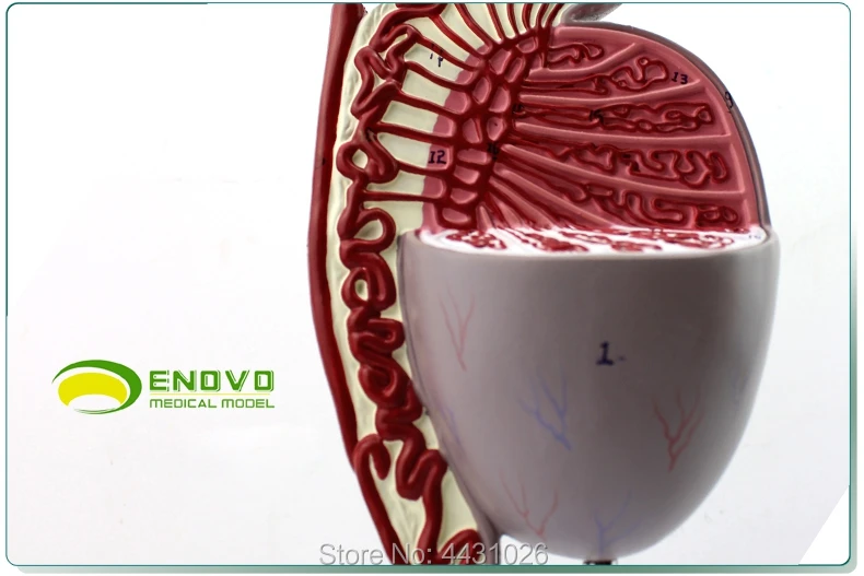 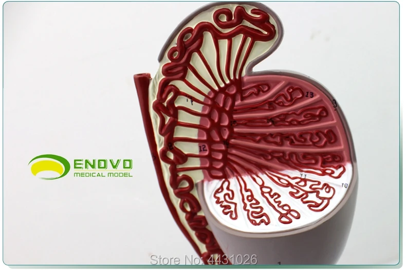 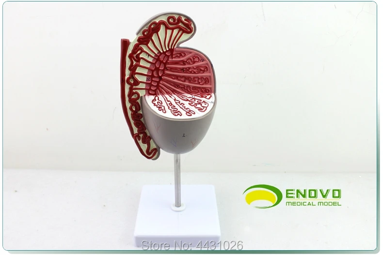 Payment 1.AliExpress supports Visa,Boleto,MasterCard,Maestro Debit Card,Western Union,Webmoney,QIWI and wire Transfer via banks.
2.If you still have any questions on the payment, Feel free to contact us.  Shipping 1.We will send the items within 2-3 days once system confirms buyers payment.
2.The shipping cost does not include any import taxes,the buyers are responsible for customs duties.
3.We will refund buyers if buyers return the items within 15 days of buyers receipt of the items for any reason. However,the buyers should bear the return shipping freight and make sure that the items returned are in their original conditions.
4.Items are shipped by AliExpress Standard Shipping , AliExpress Premium Shipping , ePacket,reach most of countries within 15 to 35 bussiness days. Delivery time depends on destination and other factors,it may take up to 60 business days. 5.If you want to get the item faster,please contact us to pay additional freight, we can offer you the following faster and cheaper shipping ways:UPS,DHL,Fedex,TNT,DEPX,EMS.Via these ways, you can get the item within about 5-7 business days.  Feedback 1.Your satisfaction and positive feedback is very important to us.Please leave positive feedback and 5 stars if you are satisfied with our items and services.
2.If you have any problems with our items or services,please feel free to contact us first before you leave negative feedback.We will do our best to solve any problems and provide you with the best customer services.  
|


 배송기간
배송기간